

Core News: New USPS stamps have Ďă¸Ű6şĎżŞ˝±˝áąű connection
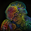
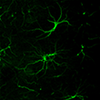
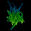
 Oak Leaf Surface, (Right) Mouse Brain Neurons")
Dec. 13, 2022 – The U.S. Postal Service announced seven new stamp subjects for 2023, and this round there is a Houston connection. Two images produced by Jason Kirk, Director of the Optical Imaging & Vital Microscopy Core at Ďă¸Ű6şĎżŞ˝±˝áąű, were chosen as part of the Life Magnified category. They are among 20 different images chosen by the USPS that capture details of life undetectable by the human eye. They are taken with microscopes and highly specialized photographic techniques that can capture the fine details found in nature and in many cases used in scientific research.
Learn more about the cornerstone of our imaging capabilities that can be used with a wide array of sample types.
LightSheet Microscopy
Learn more about how this novel 3D imaging technology can provide more insight into cleared whole mounts.
Confocal Microscopy
Learn more about how our confocal microscopes can improve your fluorescence images.
Multi-Photon Microscopy
Learn more about how our 2-Photon microscopes can help you see deeper into intact tissue.
µCT (Micro Computed Tomography)
Learn more about how ÎĽCT technology can generate 3D images from unlabeled samples.
OPT (Optical Projection Tomography)
Learn more about how our custom built OPT solutions provide fast optical sectioning for biological specimens.
